This is an old revision of the document!
Hemodynamic Correlates of Cognition in Human Infants
TECHNIQUES FOR MEASURING BLOOD OXYGENATION
Neural firing is metabolically demanding and results first in a local increase in deoxygenated
hemoglobin (deOxy-Hb), followed immediately by a compensatory sustained increase in
oxygenated hemoglobin (Oxy-Hb) and subsequent decrease in deOxy-Hb (see Figure 1). The
specific relationship of the circulatory system to local changes in neural activity is referred to as
neurovascular coupling.
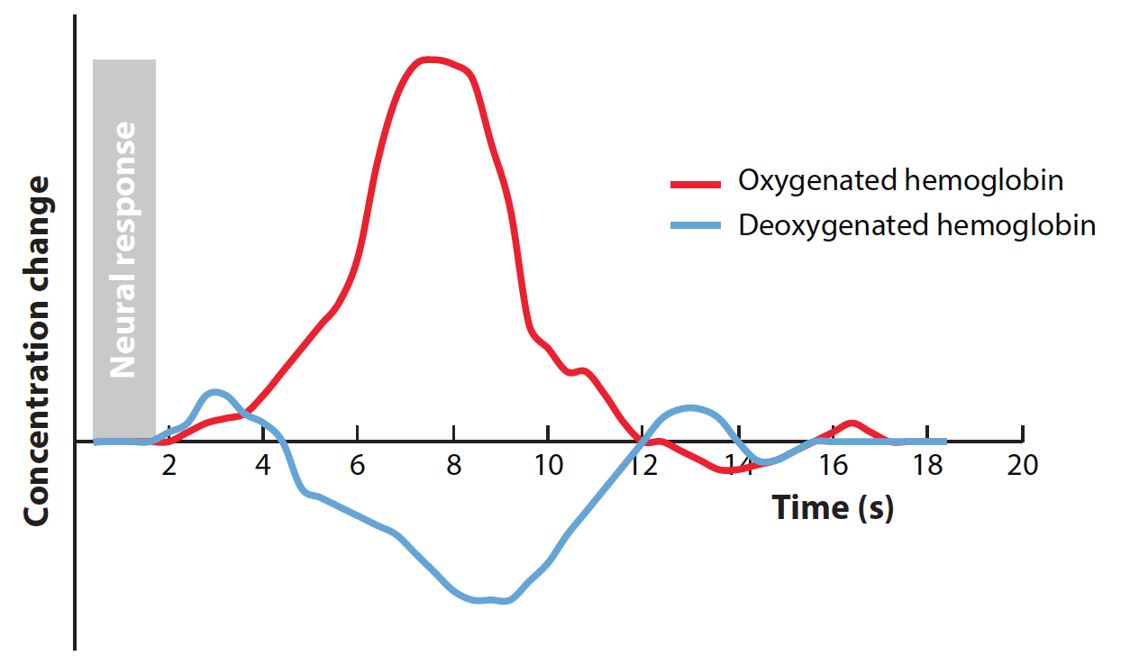
When a specific pool of neurons is active, the hemodynamic response is believed to be localized
within 1–3mmof this neural activity (e.g., Shmuel et al. 2007, using 7T in human occipital cortex,
V1) and likely represents the local field potentials rather than spiking activity of clusters of neurons
(Goense&Logothetis 2008).However, the hemodynamic response unfolds slowly compared with
the rapid change in neural activity. Even after a brief burst of stimulus-evoked neural activity
(<1 s), the adult hemodynamic response peaks approximately 6 s later and the concentrations of
Oxy-Hb and deOxy-Hb do not return to baseline until more than 10 s after the stimulus was
initially presented (Malonek & Grinvald 1996) (see Figure 1). These spatial-temporal aspects of
the hemodynamic response greatly constrain the designs and interpretations that can be drawn
from neuroimaging methods that rely on neurovascular coupling.
FMRI employs the presentation of radio wave pulses in a staticmagnetic field to record what is called the blood-oxygen-level-dependent (BOLD)
response that varies with fluctuations in local concentrations of deOxy-Hb. By contrast, fNIRS
simultaneously records changes in bothOxy-Hb and deOxy-Hb by employing two wavelengths of
near-infrared light (e.g., 690 nm and 830 nm), which span the crossover point in their respective
absorbance spectra.
While the spatial resolution of fMRI varieswith the strength of the staticmagnetic field (B0; e.g.,
1.5T, 3T, or 7T) and the specific scanning parameters, the typical fMRI voxel size of 3mm3 is well
matched to the resolution of the hemodynamic response itself. In contrast, current fNIRS systems
have a much coarser spatial resolution. As shown in Figure 2, near-infrared light is delivered to
the surface of the scalp via optical fibers (emitters). Surrounding each emitter are one or more
optical fibers (detectors) that collect a small fraction of the photons that return to the surface of
the scalp after passing through various layers of neural tissue between the emitter and the detector.
Each pair of optical fibers that link an emitter and a detector is referred to as a fNIRS channel
and samples the underlying tissue from which the hemodynamic response is being recorded.
Photons leaving the emitter and reaching the detector are primarily absorbed in the surface layers
above the cortex, including the scalp, skull, cerebral-spinal fluid, and surface vasculature (see
Figure 2). The remaining photons travel along a banana-shaped trajectory that dips into the
underlying cortex. The distance between the emitter and detector determines the lowest point of
the banana-shaped trajectory; the greater the separation between the two, the deeper the trajectory
through the cortex. However, greater separation also leads to greater attenuation of the signal.
Current systems typically employ emitter-detector separations of 2–3 cm, which optimize cortical
sampling and signal strength.However, photons must necessarily enter and exit the brain through the scalp and therefore are substantially modulated by absorption in the superficial layers of tissue
between the surface of the cortex and the scalp. This introduces a substantial source of noise into
what is a rather weak signal. Thus, signal averaging, as in the ERP, must be used to factor out
noncortical noise (under the assumption that background systemic vascular effects are uncorrelated
with the presentation of a stimulus).
One final consideration regarding spatial resolution of fNIRS is skull thickness. The thickness
of the skull is directly related to the amount of cortex that is being recorded from in a given fNIRS
channel. This is important not only because the skull is quite thin in neonates (6mmon average) and
becomes thicker with age (10 mm by age 7) but also because skull thickness varies across regions
of the head within a given age (Beauchamp et al. 2011), creating a confound when examining
absolute changes in cortical activation across ages or across brain regions (e.g., hemispheres). This
underlying anatomy should be taken into consideration for studies that compare neural activity
to a given stimulus across fNIRS channels without normalizing these activations to a second
stimulus. In the future, anatomical data that compensate for this differential path length from
fNIRS could help to prevent the false attribution of hemispheric and regional differences (unless
only relative changes in activation comprise the dependent measure). In addition to variability in
skull thickness, it is important to determine the distance from the tips of the optical fibers on the
scalp to the depth of the cortical area that is being targeted in a given recording session.